Different Between Gram-negative and Gram-positive Bacteria
Danish scientist Hans Christian Gram devised a method to differentiate two types of bacteria based on the structural differences in their cell walls. In his test, bacteria that retain the crystal violet dye do so because of a thick layer of peptidoglycan and are called Gram-positive bacteria. In contrast, Gram-negative bacteria do not retain the violet dye and are colored red or pink. Compared with Gram-positive bacteria, Gram-negative bacteria are more resistant against antibodies because of their impenetrable cell wall. These bacteria have a wide variety of applications ranging from medical treatment to industrial use and Swiss cheese production.
Comparison chart
| Gram-negative Bacteria | ||
|---|---|---|
| Gram reaction | Can be decolourized to accept counter stain (Safranin or Fuchsine); stain red or pink, they don't retain the Gram stain when washed with absolute alcohol and acetone. | Retain crystal violet dye and stain dark violet or purple, they remain coloured blue or purple with gram stain when washed with absolute alcohol and water. |
| Peptidoglycan layer | Thin (single-layered) | Thick (multilayered) |
| Teichoic acids | Absent | Present in many |
| Periplasmic space | present | Absent |
| Outer membrane | Present | Absent |
| Lipopolysaccharide (LPS) content | High | Virtually none |
| Lipid and lipoprotein content | High (due to presence of outer membrane) | Low (acid-fast bacteria have lipids linked to peptidoglycan) |
| Flagellar structure | 4 rings in basal body | 2 rings in basal body |
| Toxins produced | Primarily Endotoxins | Primarily Exotoxins |
| Resistance to physical disruption | Low | High |
| Inhibition by basic dyes | Low | High |
| Susceptibility to anionic detergents | Low | High |
| Resistance to sodium azide | Low | High |
| Resistance to drying | Low | High |
| Cell wall composition | The cell wall is 70-120 Å (ångström) thick; two layered. Lipid content is 20-30% (high), Murein content is 10-20% (low). | The cell wall is 100-120 Å thick; single layered. Lipid content of the cell wall is low , whereas Murein content is 70-80% (higher). |
| Mesosome | Mesosome is less prominent. | Mesosome is more prominent. |
| Antibiotic Resistance | More resistant to antibiotics. | More susceptible to antibiotics |
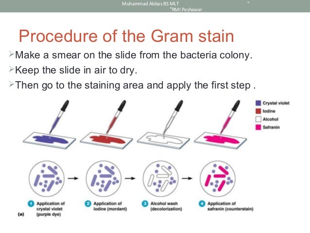
Staining and Identification

In a Gram stain test, bacteria are washed with a decolorizing solution after being dyed with crystal violet. On adding a counterstain such as safranin or fuchsine after washing, Gram-negative bacteria are stained red or pink while Gram-positive bacteria retain their crystal violet dye.
This is due to the difference in the structure of their bacterial cell wall. Gram-positive bacteria do not have an outer cell membrane found in Gram-negative bacteria. The cell wall of Gram-positive bacteria is high in peptidoglycan which is responsible for retaining the crystal violet dye.


The following videos demonstrate the staining of Gram-positive and negative bacteria respectively.
Pathogenesis in humans
Both gram-positive and gram-negative bacteria can be pathogenic (see list of pathogenic bacteria). Six gram-positive genera of bacteria are known to cause disease in humans: Streptococcus, Staphylococcus, Corynebacterium, Listeria, Bacillus and Clostridium. Another 3 cause diseases in plants: Rathybacter, Leifsonia, and Clavibacter.
Many gram-negative bacteria are also pathogenic e.g., Pseudomonas aeruginosa, Neisseria gonorrhoeae, Chlamydia trachomatis, and Yersinia pestis. Gram-negative bacteria are also more resistant to antibiotics because their outer membrane comprises a complex lipopolysaccharide (LPS) whose lipid portion acts as an endotoxin. They also develop resistance sooner:
A lot of Gram-negative bacteria, they come out of the box, if you will, resistant to a number of important antibiotics that we might use to treat them. We’re talking about agents with names like Acinetobacter, Pseudomonas, E. coli. These are bacteria that have historically done a very good job of very quickly developing resistance to antibiotics. They have a lot of tricks up their sleeves for developing resistance to antibiotics, so they’re a group of agents that can quickly become resistant, can pose major challenges to resistance. And what we’ve seen over the past decade is these Gram-negative agents becoming very rapidly more and more resistant to all of the agents that we have available to treat them.
Greater resistance of gram-negative bacteria also applies to a newly discovered class of antibiotics that was announced in early 2015 after a decades-long drought in new antibiotics. These drugs are not likely to work on gram-negative bacteria.

Gram positive Cocci
Bacteria are classified based on their cell shape into bacilli (rod shaped) and cocci (sphere shaped).Typical Gram-positive cocci stains include (pictures):
- Clusters: usually characteristic of Staphylococcus, such as S. aureus
- Chain: usually characteristic of Streptococcus, such as S. pneumoniae, B group streptococci
- Tetrad: usually characteristic of Micrococcus.
Gram-positive bacilli tend to be thick, thin or branching.
Commercial uses of non-pathogenic Gram-positive bacteria

Many streptococcal species are nonpathogenic, and form part of the commensal human microbiome of the mouth, skin, intestine, and upper respiratory tract. They are also a necessary ingredient in producing Emmentaler (Swiss) cheese.
Non-pathogenic species of corynebacterium are used in industrial production of amino acids, nucleotides, bioconversion of steroids, degradation of hydrocarbons, cheese ageing, production of enzymes etc.
Many Bacillus species are able to secrete large quantities of enzymes.
- Bacillus amyloliquefaciens is the source of a natural antibiotic protein barnase (a ribonuclease), alpha amylase used in starch hydrolysis, the protease subtilisin used with detergents, and the BamH1 restriction enzyme used in DNA research.
- C. thermocellum can utilize lignocellulose waste and generate ethanol, thus making it a possible candidate for use in production of ethanol fuel. It is anaerobicand is thermophilic, which reduces cooling cost.
- C. acetobutylicum, also known as the Weizmann organism, was first used by Chaim Weizmann to produce acetone and biobutanol from starch in 1916 for the production of gunpowder and TNT.
- C. botulinum produces a potentially lethal neurotoxin that is used in a diluted form in the drug Botox. It is also used to treat spasmodic torticollis and provides relief for approximately 12 to 16 weeks.
The anaerobic bacterium C. ljungdahlii can produce ethanol from single-carbon sources including synthesis gas, a mixture of carbon monoxide and hydrogen that can be generated from the partial combustion of either fossil fuels or biomass.
Gram-indeterminate and Gram-variable Bacteria
Not all bacteria can be reliably classified through Gram staining. For example, acid-fast bacteria or Gram-variable do not respond to Gram staining.


No comments:
Post a Comment