My Blog List

Wednesday, June 19, 2019

Media preparation and sterilization

                                                    Media Preparation and Sterilization

Definition of media

Image result for nutrient broth

A culture media is a special medium used in microbiological laboratories to grow different kinds of microorganisms. ... Different nutrients and chemicals are added to it to allow the growth of different microorganisms

Bacteria and fungi are grown on or in microbiological media of various types. The medium that is used to culture the microorganism depends on the microorganism that one is trying to isolate or identify. Different nutrients may be added to the medium, making it higher in protein or in sugar. Various pH indicators are often added for differentiation of microbes based on their biochemical reactions: the indicators may turn one color when slightly acidic, another color when slightly basic. Other added ingredients may be growth factors, NaCl, and pH buffers which keep the medium from straying too far from neutral as the microbes metabolize

Type of media

There are different type of media used in laboratory.

Since there are many types of microorganisms, each having unique properties and requiring specific nutrients for growth, there are many types based on what nutrients they contain and what function they play in the growth of microorganisms.

1 Solid media
2 Liquid media

1 Solid media. Nutrient agar media

A culture may be solid or liquid. The solid culture media is composed of a brown jelly like substance known as agar. Different nutrients and chemicals are added to it to allow the growth of different microorganisms

Nutrient agar media

Image result for nutrient broth
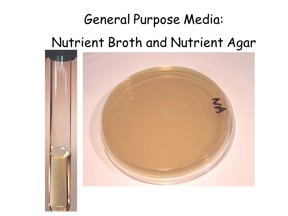
Nutrient Agar is a general purpose, nutrient medium used for the cultivation of microbes supporting growth of a wide range of non-fastidious organisms. Nutrient agar is popular because it can grow a variety of types of bacteria and fungi, and contains many nutrients needed for the bacterial growth

Source of agar           [agar is taken from the sea weed, Algae]

The gelling agent in agar is an unbranched polysaccharide obtained from the cell walls of some species of red algae, primarily from the genera Gelidium and Gracilaria. For commercial purposes, it is derived primarily from Gelidium amansii. In chemical terms, agar is a polymer made up of subunits of the sugar galactose

Composition of nutrient agar for 100ml

IngredientsQuantity (100 mL)
Peptone0.5 g
Yeast Extract0.2 g
Sodium Chloride0.5 g
Agar1.5 g
pH7

Preparation of Nutrient Agar
1. Suspend 28 g of nutrient agar powder in 1 litre of distilled water.
2. Heat this mixture while stirring to fully dissolve all components.
3. Autoclave the dissolved mixture at 121 degrees Celsius for 15 minutes.
4. Once the nutrient agar has been autoclaved, allow it to cool but not solidify.
5. Pour nutrient agar into each plate and leave plates on the sterile surface until the agar has solidified.
6. Replace the lid of each Petri dish and store the plates in a refrigerator.
Uses of Nutrients Agar
1. It is frequently used for isolation and purification of cultures.
2. It can also be used as a means for producing the bacterial lawns needed for antibiotic sensitivity tests.  In actuality, antibiotic sensitivity testing is typically performed on media specially formulated for that purpose.

2. Liquid media

This is a liquid medium which allows the microorganisms to multiply and has the essential nutrients that are required for it. It is usually composed of bacteria taken from a liquid source such as pond water. The basic nutrient broth is the most commonly used. Selective Media Plate

Nutrient Broth. 

Image result for nutrient broth

Nutrient broth is a basic media composed of a simple peptone and a beef extract. Peptone contributes organic nitrogen in the form of amino acids and long-chained fatty acids. Beef Extract provides additional vitamins, carbohydrates, salts and other organic nitrogen compounds

Composition of nutrient broth media

Nutrient broth. Add 13 g of nutrient broth powder to 1 litre of distilled water

aa Amount in  in 100gm


Weigh 1.3 gram of nutrient broth powder and put it in a conical flask with a cotton plug. Measure 100 ml of distilled water in a measuring cylinder and add in to conical flask containing nutrient broth powder. Dissolve the nutrient broth powder in the water by swirling. Autoclave it at 15 psi pressure in autoclave

Composition of nutrient broth for one liter water

Intended use Nutrient Broth is used for the general cultivation of less fastidious microorganisms , can be enriched with blood or other biological fluids.
 Composition** 
Ingredients                                                                  Gms / Litre 

Peptone                                                                           5.000
 Sodium chloride                                                             5.000
 HM peptone B#                                                              1.500
 Yeast extract                                                                   1.500 Final 
pH                                                                                 ( at 25°C) 7.4±0.2


 **Formula adjusted, standardized to suit performance parameters Directions Suspend 13.0 grams in 1000 ml distilled water. Heat, if necessary, to dissolve the medium completely. Dispense into tubes or flasks as desired. Sterilize by autoclaving at 15 lbs pressure (121°C) for 15 minutes

Friday, June 14, 2019

Overview of the Immune System


                                Overview of the Immune System

Image result for innate immunity

The immune system is the body's defense against infectious organisms and other invaders The immune system is made up of a network of cells, tissues, and organs that work together to protect the body,it is also help the infection of any pathogens
CST supports your immunology research with the highest quality antibodies. Immunology-based research focuses on the components of the human immune system and the possibility of their use to combat various diseases.
You need tools to identify and analyze immune cells and immune signaling to advance your research. CST is proud to produce top of the line antibodies for investigating proteins within the complex signaling pathways of the immune system and to contribute to the international movement to develop better, more efficacious therapeutics.
The immune system is comprised of two broad cellular responses:
  • Innate immunity
  • Adaptive immunity

Innate Immunity

The innate immune response is your first line of defense against pathogens. It provides a quick response to pathogens by many mechanisms, including cytokine production and complement activation.
The cell types involved in the innate immune response are phagocytic cells: neutrophils, macrophages, natural killer cells, basophils, and others.
As a leader in antibody development, CST has developed a wide array of highly effective antibodies against proteins involved in innate immunity such as the STINGNFκB, and inflammasome signaling pathways

Adaptive Immunity

The adaptive immune response uses antigen-specific receptors to detect foreign antigens. This is a slow occurrence that results from efforts of T Cells, B cells, and natural killer T Cells. Humoral immunity uses antibodies for detection, whereas cell-mediated immunity uses T Cells to destroy the affected cells.
When the antigen is encountered for the first time, lymphocytes exert the primary immune response. The same cells can “learn” from their experience, so that a subsequent encounter with the same antigen will result in a quicker, secondary immune response.
CST has developed a wide array of highly effective antibodies to label the many proteins involved in the lymphocyte signaling pathways, including the TCR and BCR.

Immune Cell Signaling

The regulation of immune cells occurs through a number of key signaling pathways. Each pathway is comprised of a complex network of proteins that interact with one another to induce a specific cellular response to stimuli.
In addition to the STING, NFƘB, inflammasome, TCR, and BCR signaling pathways, the JAK/STATand TLR signaling pathways also play major roles in immune cell signaling

Development of T Lymphocytes

T Cells play a critical role in cell-mediated immunity and arise from lymphoid progenitor cells that originally developed from hematopoietic stem cells in the bone marrow.
T Cells are identified by the expression of CD3 and develop into their various subtypes in the thymus (hence the name T Cell). Each subtype is defined by the specific receptors expressed on the cell surface, making these cells highly selective to non-self pathogens. Mature T Cells are released into the bloodstream where they can be induced to become one of several classes of T cells including:
  • Helper T Cell - CD4+ T Cells suited to recognize peptide antigens bound to class II MHC proteins and release a variety of cytokines.
  • Cytotoxic T Cell - CD8+ cells that recognize virus-infected cells or tumor cells.
  • Regulatory T Cell - The main job of regulatory T Cells (Tregs) is to maintain tolerance to self-antigens, as well as limit T-effector cell function and proliferation.
  • Natural Killer T Cell - NK cells release small granules containing granzymes and perforin, which form pores and break down intracellular proteins in order to induce apoptosis in virally-infected or tumor cells.

T Cell Activation & T Cell Receptor (TCR) Signaling

T Cells become activated when a pathogenic antigen binds their cell-surface receptors. Each cell-surface receptor has affinity to a unique antigen, making T Cells highly specialized in antigen recognition.
Once the T Cell receptors bind an antigen, the T Cell will activate a series of internal signaling pathways that allow for the antigen recognition to be verified. Only then will the T cell proliferate, expanding the pool of available cells that are specific for the harmful antigen, such as to different bacteria and parasites.
CST has a wide array of expertly validated antibodies that can distinguish T cell populations accurately and reproducibly by WB, IF/IHC, IP and extracellular/intracellular flow cytometry.

T Cell Immunophenotyping

Using specific antibodies, individual subtypes of T Cells can be distinguished from a heterogeneous pool of immune cells. This type of immune cell subtyping is known as immunophenotyping.
Immunophenotyping of T Cells can be accomplished because T Cells have specific cell surface receptors, intracellular cytokine expression profiles, and/or differential phosphorylation of other intercellular proteins. This allow for selective labeling of these cells with specific markers. T Cells can first be immunophenotyped using IHC methods based on their CD3 expression, then they can be further sub-classified based on other well-characterized markers.

Development of B Lymphocytes

B Cells differentiate from hematopoietic cells found in the bone marrow. Immunoglobulin (Ig) receptors are assembled on the surface of B Cells and allow for specific recognition of a single antigen. Varying gene rearrangements during development lead to the differential expression of Ig surface receptors. Mature B Cells migrate to the lymph nodes and spleen after which they can further become memory B Cells or plasma cells.
While T Cells and B Cells are both able to recognize pathogens such as bacteria, viruses, and apoptotic cells, there two cell types are distinct in their characteristics and functions. B Cells develop in the bone marrow while T Cells are formed in the thymus.
The main difference is that T Cells can only recognize viral antigens outside the infected cells whereas B Cells can recognize the surface antigens of bacteria and viruses, which in turn results in antibody production.

B Lymphocyte Activation and Antibody Production

The critical cells that mediate antibody production are B Cells. Specific B cell receptors recognize and facilitate processing of antigens. B Cell activation occurs with help from T Cells, allowing the B Cells to mature into plasma cells that secrete antibodies.

B Cell Signaling

The B Cell receptor (BCR) binds antigens, resulting in activation of multiple signaling cascades that involve kinases, GTPases, and transcription factors. This results in changes in cell metabolism, gene expression, and cytoskeletal organization. The complexity of BCR signaling permits many distinct outcomes including survival, apoptosis, proliferation, or differentiation into plasma cells or memory B Cells.
CST has a wide array of expertly validated antibodies that can distinguish B cell populations accurately and reproducibly by WB, IF/IHC, IP and extracellular/intracellular flow cytometry.
  B Cell Immuophenoty Through out the various stages of B Cell development and maturation, B Cells express many different markers that can be distinguished by IHC or flow cytometry. Using specific antibodies, stages of individual B Cells can be distinguished from a heterogeneous pool of immune cells. This type of immune cell subtyping is known as immunophenotyping.
CST is your one-stop shop for B Cell immunophenotyping. CST has a wide array of expertly validated antibodies that can distinguish B Cell populations accurately and reproducibly by IHC including:

Thursday, June 13, 2019

Bright-Field Light Microscope and Microscopic Measurement of Organisms SAFETY and CONSIDERATIONS


Bright-Field Light Microscope and Microscopic Measurement of Organisms SAFETY and CONSIDERATIONS

Related image

Slides and coverslips are glass. Be careful with them. Do
not cut yourself when using them. The coverslips are
very thin and easily broken. Dispose of any broken glass
in the appropriately labeled container. If your micro
scope has an automatic stop, do not use it as the stage
micrometer is too thick to allow it to function properly.
It may result in a shattered or broken slide or lens


Medical Application

In the clinical laboratory, natural cell size, arrangement and
motility are important characteristics in the identification of
a bacterial pathogen


Materials per Studentcompound microscope
lens paper and lens cleaner
immersion oil
prepared stained slides of several types of bacteria
(rods, cocci, spirilla), fungi, algae, and protozoa
glass slides
coverslips
dropper with bulb
newspaper or cut-out letter
e’s
tweezers
ocular micrometer
stage micrometer
Learning ObjectivesEach student should be able to
1. Identify all the parts of a compound microscope
2. Know how to correctly use the microscope—
especially the oil immersion lens
3. Learn how to make and examine a wet-mount
preparation
4. Understand how microorganisms can be measured
under the light microscope
5. Calibrate an ocular micrometer
6. Perform some measurements on different
microorganisms

Why Are Prepared Slides

Used in This Exercise?Because this is a microbiology course and most of the microorganisms studied are bacteria, this is an excellent place
to introduce the student to the three basic bacterial shapes:
cocci, rods, and spirilla. By gaining expertise in using the
bright-field light microscope, the student should be able to
observe these three bacterial shapes by the end of the lab
period. In addition, the student will gain an appreciation for
the small size and arrangement of procaryotic cell structure.
One major objective of this exercise is for the student
to understand how microorganisms can be measured under
the light microscope and to actually perform some measurements on different microorganisms. By making measurements on prepared slides of various bacteria, fungi,
algae, and protozoa, the student will gain an appreciation
for the size of different microorganisms discussed throughout both the lecture and laboratory portions of this course

Principles

The bright-field light microscope is an instrument
that magnifies images using two lens systems. Initial
magnification occurs in the
objective lens. Most microscopes have at least three objective lenses on a rotating base, and each lens may be rotated into alignment with the eyepiece or ocular lens in which the
final magnification occurs. The objective lenses are
identified as the
low-power, high-dry, and oil immersion objectives. Each objective is also designated by
other terms. These terms give either the
linear magnification or the focal length. The latter is about equal
to or greater than the
working distance between the
specimen when in focus and the tip of the objective
lens. For example, the low-power objective is also
called the
10×, or 16 millimeter (mm), objective; the
high-dry is called the
40×, or 4 mm, objective; and
the oil immersion is called the
90×, 100×, or 1.8 mm
objective.
As the magnification increases, the size of
the lens at the tip of the objective becomes progressively smaller and admits less light. This is one of the
reasons that changes in position of the
substage condenser and iris diaphragm are required when using
different objectives if the specimens viewed are to be
seen distinctly. The condenser focuses the light on a
small area above the stage, and the iris diaphragm controls the amount of light that enters the condenser.

Immersion lens


When the oil immersion lens is used, immersion oil
fills the space between the objective and the specimen.
Because immersion oil has the same
refractive indexas glass, the loss of light is minimized (figure 1.1). Theeyepiece, or ocular, at the top of the tube magnifies
the image formed by the objective lens. As a result, the
total magnification seen by the observer is obtained by
multiplying the magnification of the objective lens by
the magnification of the ocular, or eyepiece. For example, when using the 10
× ocular and the 43× objective,
total magnification is 10
× 43 = 430 times.

Procedure for Basic Microscopy:Proper Useof the Microscope

1. Always carry the microscope with two hands. Place
it on the desk with the open part away from you.
2. Clean all of the microscope’s lenses only with
lens paper and lens cleaner if necessary. Do not
use paper towels or Kimwipes; they can scratch
the lenses. Do not remove the oculars or any other
parts from the body of the microscope.
3. Cut a lowercase e from a newspaper or other
printed page. Prepare a wet-mount as illustrated in
figure 1.2. Place the glass slide on the stage of the
microscope and secure it firmly using stage clips.
If your microscope has a mechanical stage device,
place the slide securely in it. Move the slide until
the letter
e is over the opening in the stage.
4. With the low-power objective in position, lower
the tube until the tip of the objective is within
5 mm of the slide. Be sure that you lower the tube
while looking at the microscope from the side.
5. Look into the microscope and slowly raise the
tube by turning the coarse adjustment knob
counterclockwise until the object comes into
view. Once the object is in view, use the fine
adjustment knob to focus the desired image.
6. Open and close the diaphragm, and lower and raise
the condenser, noting what effect these actions
have on the appearance of the object being viewed.
Usually the microscope is used with the substage
condenser in its topmost position. The diaphragm
should be open and then closed down until just a
slight increase in contrast is observed (table 1.1).
7. Use the oil immersion lens to examine the stained
bacteria that are provided (figure 1.3
a–d). The
directions for using this lens are as follows: First locate

Examples of Bacterial Shapes as Seen with the Bright-field Light Microscope.


(a) Staphylococcus aureus cocci; singular,
coccus (
×1,000). 
Image result for staphylococcus aureus

(b) Bacillus subtilis rods or bacilli; singular, bacillus (×1,000).

Image result for bacillus subtilis
 (c) A single, large spirillum; plural, spiralla (Spirillum volutans;×1,000).
Image result for spirillum bacteria

 (d) Numerous, small spirilla (Rhodospirillum rubrum; ×1,000).
Image result for rhodospirillum bacteria

Table 1.1 Troubleshooting the Bright-Field Light Microscope

Common Problem Possible Correction
No light passing through the ocular Check to ensure that the microscope is completely plugged into a good receptacleCheck to ensure that the power switch to the microscope is turned onMake sure the objective is locked or clicked in placeMake sure the iris diaphragm is openInsufficient light passing through the ocular Raise the condenser as high as possibleOpen the iris diaphragm completelyMake sure the objective is locked or clicked in placeLint, dust, eyelashes interferring with view Clean ocular with lens paper and cleanerParticles seem to move in hazy visual field Air bubbles in immersion oil; add more oil or make certain that oil immersion objective is in the oilMake sure that the high-dry objective is not being used with oilMake sure a temporary coverslip is not being used with oil. Oil causes the coverslip to float since the coverslipsticks to the oil and not the slide, making viewing very hazy or impossible

Mechanism of Action of Hydroxychloroquine as an Antirheumatic Drug

Mechanism of Action of Hydroxychloroquine as an Antirheumatic Drug Mechanism of Action of Hydroxychloroquine as an Antirheumat...