My Blog List

Monday, April 29, 2019

A Brief History of Microbiology And modern microbiology


A Brief History of Microbiology


 Microbiology has had a long, rich history, initially centered in the causes of infectious diseases but now including practical applications of the science. Many individuals have made significant contributions to the development of microbiology.
Early history of microbiology. Historians are unsure who made the first observations of microorganisms, but the microscope was available during the mid1600s, and an English scientist namedRobert Hooke made key observations. He is reputed to have observed strands of fungi among the specimens of cells he viewed. In the 1670s and the decades thereafter, a Dutch merchant namedAnton van Leeuwenhoek made careful observations of microscopic organisms, which he called animalcules. Until his death in 1723, van Leeuwenhoek revealed the microscopic world to scientists of the day and is regarded as one of the first to provide accurate descriptions of protozoa, fungi, and bacteria.
After van Leeuwenhoek died, the study of microbiology did not develop rapidly because microscopes were rare and the interest in microorganisms was not high. In those years, scientists debated the theory of spontaneous generation, which stated that microorganisms arise from lifeless matter such as beef broth. This theory was disputed by Francesco Redi, who showed that fly maggots do not arise from decaying meat (as others believed) if the meat is covered to prevent the entry of flies. An English cleric named John Needham advanced spontaneous generation, but Lazzaro Spallanzanidisputed the theory by showing that boiled broth would not give rise to microscopic forms of life.
Louis Pasteur and the germ theory. Louis Pasteur worked in the middle and late 1800s. He performed numerous experiments to discover why wine and dairy products became sour, and he found that bacteria were to blame. Pasteur called attention to the importance of microorganisms in everyday life and stirred scientists to think that if bacteria could make the wine “sick,” then perhaps they could cause human illness.
Pasteur had to disprove spontaneous generation to sustain his theory, and he therefore devised a series of swannecked flasks filled with broth. He left the flasks of broth open to the air, but the flasks had a curve in the neck so that microorganisms would fall into the neck, not the broth. The flasks did not become contaminated (as he predicted they would not), and Pasteur's experiments put to rest the notion of spontaneous generation. His work also encouraged the belief that microorganisms were in the air and could cause disease. Pasteur postulated the germ theory of disease, which states that microorganisms are the causes of infectious disease.
Pasteur's attempts to prove the germ theory were unsuccessful. However, the German scientist Robert Koch provided the proof by cultivating anthrax bacteria apart from any other type of organism. He then injected pure cultures of the bacilli into mice and showed that the bacilli invariably caused anthrax. The procedures used by Koch came to be known as Koch's postulates (Figure ). They provided a set of principles whereby other microorganisms could be related to other diseases.
The development of microbiology. In the late 1800s and for the first decade of the 1900s, scientists seized the opportunity to further develop the germ theory of disease as enunciated by Pasteur and proved by Koch. There emerged a Golden Age of Microbiology during which many agents of different infectious diseases were identified. Many of the etiologic agents of microbial disease were discovered during that period, leading to the ability to halt epidemics by interrupting the spread of microorganisms.
Despite the advances in microbiology, it was rarely possible to render lifesaving therapy to an infected patient. Then, after World War II, the antibiotics were introduced to medicine. The incidence of pneumonia, tuberculosis, meningitis, syphilis, and many other diseases declined with the use of antibiotics.
Work with viruses could not be effectively performed until instruments were developed to help scientists see these disease agents. In the 1940s, theelectron microscope was developed and perfected. In that decade, cultivation methods for viruses were also introduced, and the knowledge of viruses developed rapidly. With the development of vaccines in the 1950s and 1960s, such viral diseases as polio, measles, mumps, and rubella came under control.
Modern microbiology. Modern microbiology reaches into many fields of human endeavor, including the development of pharmaceutical products, the use of qualitycontrol methods in food and dairy product production, the control of diseasecausing microorganisms in consumable waters, and the industrial applications of microorganisms. Microorganisms are used to produce vitamins, amino acids, enzymes, and growth supplements. They manufacture many foods, including fermented dairy products (sour cream, yogurt, and buttermilk), as well as other fermented foods such as pickles, sauerkraut, breads, and alcoholic beverages.
One of the major areas of applied microbiology is biotechnology. In this discipline, microorganisms are used as living factories to produce pharmaceuticals that otherwise could not be manufactured. These substances include the human hormone insulin, the antiviral substance interferon, numerous bloodclotting factors and clotdissolving enzymes, and a number of vaccines. Bacteria can be reengineered to increase plant resistance to insects and frost, and biotechnology will represent a major application of microorganisms in the next century.
Image result for koch postulate

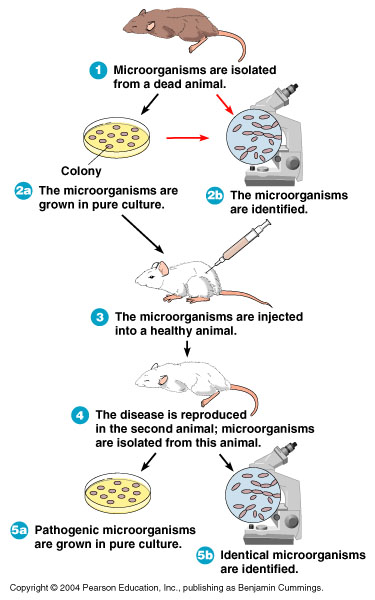
The steps of Koch's postulates used to relate a specific microorganism to a specific disease. (a) Microorganisms are observed in a sick animal and (b) cultivated in the lab. (c) The organisms are injected into a healthy animal, and (d) the animal develops the disease. (e) The organisms are observed in the sick animal and (f) reisolated in the lab.

Monday, April 22, 2019

Alexa-What’s My Sugar Level? Amazon’s Alexa is Now HIPAA Compliant

Alexa-What’s My Sugar Level? Amazon’s Alexa is Now HIPAA Compliant
Written on 04/022/2019
Vinod kushwaha


Amazon’s Alexa is Now HIPAA Compliant
Alexa is moving into health care. After a trial of Amazon speakers at patients’ rooms in Cedars-Sinai Hospitals in Los Angeles, the company this morning announced a program enabling select programmers to create and launch HIPAA-compliant health care skills that can be incorporated in Alexa. New Alex add-on abilities allow customers to ask Alexa assistant for help with matters like booking an appointment, obtaining hospital post-discharge instructions, checking on the status of more and prescription delivery.
Amazon stated the program will only allow only picked entities and business associates subject to HIPAA (the U.S. Health Insurance Portability and Accountability Act of 1996) to make these abilities for Alexa. It itself provides the environment for skill building, while the programmers are required to comply with the laws. Voice program developers who follow HIPAA guidelines now will be able to create abilities for Alexa, this is a substantial step for Amazon.
Referring to CNBC reports dated last year May, Amazon was building a healthcare team to make the voice helper useful in the health care industry. This included working through the HIPAA regulations which would be asked to achieve that.
Additionally, Amazon itself is entering healthcare alongside JP Morgan Chase and Berkshire Hathaway, which were teamed up to take on rising healthcare costs for workers. Amazon year that was last obtained online pharmacy PillPack for less than $1 billion. Along with its own capabilities that are HIPAA-compliant are enlarging. Amazon also introduced a new machine learning tool – Amazon Comprehend Medical that aggregates information using doctors’ notes & One’s health records.
Now, Amazon Alexa is supplying its “HIPAA eligible environment” to voice app developers on an invite-only foundation in the U.S. but says it hopes to enable more developers to access this capacity later on.
Developers will be able to use the Alexa Skills Kit, which supports abilities that are able to transmit and receive protected health information.
This expansion of Amazon’s Alexa to health care is very likely to raise questions. While it’s one thing to allow Alexa to play some music or to turn on your lights, whereas enabling access for Voice assistants to one’s medical records is a giant leap. Consumers will need to know how Amazon is securing their data before they feel comfortable using Alexa’s health care abilities.
As per Techcrunch, Amazon told them it uses several layers of safety to all skill data, such as encryption, access controls and safely storing information in the Amazon cloud. HIPAA,on the other hand, has its own set of safety measures in place that includes identifying protected health information (PHI) and controlling and auditing access to PHI.
Amazon now is launching six skills that demonstrate the potential of all healthcare-related skills. These come from payors healthcare providers, pharmacy benefit managers, and health coaching businesses.
One such skill from Cigna, for example, allows qualified employees to manage their health improvement goals and earn wellness incentives; another from Livongo allows members inquire Alexa for their final blood glucose reading; parents and caregivers may give their care teams updates at Boston Hospital’s ERAS (Improved Recovery After Surgery) program.
The health care skill publishers are enthusiastic about the ability to achieve their customers through voice technology.
Amazon launched a site for its new health care abilities, which supplies a sign-up form for those who want to get”updates”. The form also includes a location to describe cases are used by the health care ability you have in mind — significance Amazon is currently using this to vet the next round of programmers to invite into the app.

Wednesday, April 3, 2019

LIST OF GRAM NEGATIVE BACTERIA

Gram-negative Bacteria


Gram-negative bacteria are bacteria that do not retain the crystal violet dye in the Gram stain protocol. Gram-negative bacteria will thus appear red or pink following a Gram stain procedure due to the effects of the counterstain (for example safranin).

The Gram Stain

In microbiology, the visualization of bacteria at the microscopic level is facilitated by the use of stains, which react with components present in some cells but not others. This technique is used to classify bacteria as either Gram-positive or Gram-negative depending on their colour following a specific staining procedure originally developed by Hans Christian Gram. Gram-positive bacteria appear dark blue or violet due to the crystal violet stain following the Gram stain procedure; Gram-negative bacteria, which cannot retain the crystal violet stain, appear red or pink due to the counterstain (usually safranin).
The reason bacteria are either Gram-positive or Gram-negative is due to the structure of their cell envelope. (The cell envelope is defined as the cell membrane and cell wall plus an outer membrane, if one is present.) Gram-positive bacteria, for example, retain the crystal violet due to the amount of peptidoglycan in the cell wall. It can be said therefore that the Gram-stain procedure separates bacteria into two broad categories based on structural differences in the cell envelope.
Image result for gram staining procedure

Cell envelope of Gram-negative Bacteria

The Gram negative cell envelope contains an additional outer membrane composed by phospholipids and lipopolysaccharides which face the external environment. The highly charged nature of lipopolysaccharides confer an overall negative charge to the Gram negative cell wall. The chemical structure of the outer membrane lipopolysaccharides is often unique to specific bacterial strains (i.e. sub-species) and is responsible for many of the antigenic properties of these strains. Many species of Gram-negative bacteria are pathogenic. This pathogenicity is often associated with the lipopolysaccharide (LPS) layer of the Gram-negative cell envelope.

Mycobacterium

Mycobacterium is a genus of Actinobacteria, given its own family, the Mycobacteriaceae. Mycobacteria have a cell envelope which is not typical of Gram positives or Gram negatives. The mycobacterial cell envelope does not consist of the outer membrane characteristic of Gram negative bacteria, but has a significant peptidoglycan-arabinogalactan-mycolic acid wall structure which provides an external permeability barrier.

Characteristics of Gram-negative Bacteria

Gram-negative bacteria have a characteristic cell envelope structure very different from Gram-positive bacteria. Gram-negative bacteria have a cytoplasmic membrane, a thin peptidoglycan layer, and an outer membrane containing lipopolysaccharide. There is a space between the cytoplasmic membrane and the outer membrane called the periplasmic space or periplasm. The periplasmic space contains the loose network of peptidoglycan chains referred to as the peptidoglycan layer.

 Gram-negative bacteria

Different Between Gram-negative and Gram-positive Bacteria with Comparison chart


Different Between Gram-negative and Gram-positive Bacteria 
Danish scientist Hans Christian Gram devised a method to differentiate two types of bacteria based on the structural differences in their cell walls. In his test, bacteria that retain the crystal violet dye do so because of a thick layer of peptidoglycan and are called Gram-positive bacteria. In contrast, Gram-negative bacteria do not retain the violet dye and are colored red or pink. Compared with Gram-positive bacteria, Gram-negative bacteria are more resistant against antibodies because of their impenetrable cell wall. These bacteria have a wide variety of applications ranging from medical treatment to industrial use and Swiss cheese production.

Comparison chart

Gram-negative Bacteria versus Gram-positive Bacteria comparison chart
Gram-negative Bacteria
Gram-positive Bacteria
Gram reactionCan be decolourized to accept counter stain (Safranin or Fuchsine); stain red or pink, they don't retain the Gram stain when washed with absolute alcohol and acetone.Retain crystal violet dye and stain dark violet or purple, they remain coloured blue or purple with gram stain when washed with absolute alcohol and water.
Peptidoglycan layerThin (single-layered)Thick (multilayered)
Teichoic acidsAbsentPresent in many
Periplasmic spacepresentAbsent
Outer membranePresentAbsent
Lipopolysaccharide (LPS) contentHighVirtually none
Lipid and lipoprotein contentHigh (due to presence of outer membrane)Low (acid-fast bacteria have lipids linked to peptidoglycan)
Flagellar structure4 rings in basal body2 rings in basal body
Toxins producedPrimarily EndotoxinsPrimarily Exotoxins
Resistance to physical disruptionLowHigh
Inhibition by basic dyesLowHigh
Susceptibility to anionic detergentsLowHigh
Resistance to sodium azideLowHigh
Resistance to dryingLowHigh
Cell wall compositionThe cell wall is 70-120 Å (ångström) thick; two layered. Lipid content is 20-30% (high), Murein content is 10-20% (low).The cell wall is 100-120 Å thick; single layered. Lipid content of the cell wall is low , whereas Murein content is 70-80% (higher).
MesosomeMesosome is less prominent.Mesosome is more prominent.
Antibiotic ResistanceMore resistant to antibiotics.More susceptible to antibiotics


Staining and Identification

Microscopic view of dental plaque, showing Gram-positive (purple) and negative (red) bacteria
magnify
Microscopic view of dental plaque, showing Gram-positive (purple) and negative (red) bacteria
In a Gram stain test, bacteria are washed with a decolorizing solution after being dyed with crystal violet. On adding a counterstain such as safranin or fuchsine after washing, Gram-negative bacteria are stained red or pink while Gram-positive bacteria retain their crystal violet dye.
This is due to the difference in the structure of their bacterial cell wall. Gram-positive bacteria do not have an outer cell membrane found in Gram-negative bacteria. The cell wall of Gram-positive bacteria is high in peptidoglycan which is responsible for retaining the crystal violet dye.
Image result for gram staining procedure

Gram-positive and negative bacteria are chiefly differentiated by their cell wall structure


Image result for gram staining procedure
magnify
Gram-positive and negative bacteria are chiefly differentiated by their cell wall structure
The following videos demonstrate the staining of Gram-positive and negative bacteria respectively.

Pathogenesis in humans

Both gram-positive and gram-negative bacteria can be pathogenic (see list of pathogenic bacteria). Six gram-positive genera of bacteria are known to cause disease in humans: Streptococcus, Staphylococcus, Corynebacterium, Listeria, Bacillus and Clostridium. Another 3 cause diseases in plants: Rathybacter, Leifsonia, and Clavibacter.
Many gram-negative bacteria are also pathogenic e.g., Pseudomonas aeruginosa, Neisseria gonorrhoeae, Chlamydia trachomatis, and Yersinia pestis. Gram-negative bacteria are also more resistant to antibiotics because their outer membrane comprises a complex lipopolysaccharide (LPS) whose lipid portion acts as an endotoxin. They also develop resistance sooner:
A lot of Gram-negative bacteria, they come out of the box, if you will, resistant to a number of important antibiotics that we might use to treat them. We’re talking about agents with names like Acinetobacter, Pseudomonas, E. coli. These are bacteria that have historically done a very good job of very quickly developing resistance to antibiotics. They have a lot of tricks up their sleeves for developing resistance to antibiotics, so they’re a group of agents that can quickly become resistant, can pose major challenges to resistance. And what we’ve seen over the past decade is these Gram-negative agents becoming very rapidly more and more resistant to all of the agents that we have available to treat them.
Greater resistance of gram-negative bacteria also applies to a newly discovered class of antibiotics that was announced in early 2015 after a decades-long drought in new antibiotics. These drugs are not likely to work on gram-negative bacteria.
Structure of a gram-positive bacterial cell.
magnify
Structure of a gram-positive bacterial cell.

Gram positive Cocci

Bacteria are classified based on their cell shape into bacilli (rod shaped) and cocci (sphere shaped).Typical Gram-positive cocci stains include (pictures):
  • Clusters: usually characteristic of Staphylococcus, such as S. aureus
  • Chain: usually characteristic of Streptococcus, such as S. pneumoniae, B group streptococci
  • Tetrad: usually characteristic of Micrococcus.
Gram-positive bacilli tend to be thick, thin or branching.

Commercial uses of non-pathogenic Gram-positive bacteria

Many streptococcal species are nonpathogenic, and form part of the commensal human microbiome of the mouth, skin, intestine, and upper respiratory tract. They are also a necessary ingredient in producing Emmentaler (Swiss) cheese.
Non-pathogenic species of corynebacterium are used in industrial production of amino acids, nucleotides, bioconversion of steroids, degradation of hydrocarbons, cheese ageing, production of enzymes etc.
Many Bacillus species are able to secrete large quantities of enzymes.
  • Bacillus amyloliquefaciens is the source of a natural antibiotic protein barnase (a ribonuclease), alpha amylase used in starch hydrolysis, the protease subtilisin used with detergents, and the BamH1 restriction enzyme used in DNA research.
  • C. thermocellum can utilize lignocellulose waste and generate ethanol, thus making it a possible candidate for use in production of ethanol fuel. It is anaerobicand is thermophilic, which reduces cooling cost.
  • C. acetobutylicum, also known as the Weizmann organism, was first used by Chaim Weizmann to produce acetone and biobutanol from starch in 1916 for the production of gunpowder and TNT.
  • C. botulinum produces a potentially lethal neurotoxin that is used in a diluted form in the drug Botox. It is also used to treat spasmodic torticollis and provides relief for approximately 12 to 16 weeks.
The anaerobic bacterium C. ljungdahlii can produce ethanol from single-carbon sources including synthesis gas, a mixture of carbon monoxide and hydrogen that can be generated from the partial combustion of either fossil fuels or biomass.

Gram-indeterminate and Gram-variable Bacteria

Not all bacteria can be reliably classified through Gram staining. For example, acid-fast bacteria or Gram-variable do not respond to Gram staining.

The Study Of Microorganisms

The Study Of Microorganisms

As is the case in many sciences, the study of microorganisms can be divided into two generalized and sometimes overlapping categories. Whereas basic microbiology addresses questions regarding the biologyof microorganisms, applied microbiology refers to the use of microorganisms to accomplish specific objectives.

Basic microbiology

The study of the biology of microorganisms requires the use of many different procedures as well as special equipment. The biological characteristics of microorganisms can be summarized under the following categories: morphologynutritionphysiologyreproductionand growthmetabolism, pathogenesis, antigenicity, and genetic properties.


MORPHOLOGY

Morphology refers to the size, shape, and arrangement of cells. The observation of microbial cells requires not only the use of microscopesbut also the preparation of the cells in a manner appropriate for the particular kind of microscopy. During the first decades of the 20th century, the compound light microscope was the instrument commonly used in microbiology. Light microscopes have a usual magnification factor of 1000 × and a maximum useful magnification of approximately 2000 ×. Specimens can be observed either after they have been stained by one of several techniques to highlight some morphological characteristics or in living, unstained preparations as a “wet mount.”

Light microscopy

Several modifications of light microscopy are available, such as:
  • bright field
    The specimen is usually stained and observed while illuminated; useful for observation of the gross morphological features of bacteria, fungi, algae, and protozoa.
  • dark field
    The specimen is suspended in a liquid on a special slide and can be observed in a living condition; useful for determining motility of microorganisms or some special morphological characteristic such as spiral or coiled shapes.
  • fluorescence
    The specimen is stained with a fluorescent dye and then illuminated; objects that take up the fluorescent dye will “glow.”
  • phase contrast
    Special condenser lenses allow observation of living cells and differentiation of cellular structures of varying density.

Electron microscopy

The development of the electron microscope and complimentary techniques vastly increased the resolving power beyond that attainable with light microscopy. This increase is possible because the wavelengths of the electron beams are so much shorter than the wavelengths of light. Objects as small as 0.02 nm are resolvable by electron microscopy, compared with 0.25 μm—allowing, for instance, the observation of virions and viral structures. Specimens are observed by either transmission electron microscopy or scanning electron microscopy. In TEM the electron beam passes through the specimen and registers on a screen forming the image; in SEM the electron beam moves back and forth over the surface of microorganisms coated with a thin film of metal and registers a three-dimensional picture on the screen.


Advances in microscopes and microscopic techniques continue to be introduced to study cells, molecules, and even atoms. Among these are confocal microscopy, the atomic force microscope, the scanning tunneling microscope, and immunoelectron microscopy. These are particularly significant for studies of microorganisms at the molecular level.

Nutritional and physiological characteristics

Microorganisms as a group exhibit great diversity in their nutritionalrequirements and in the environmental conditions that will support their growth. No other group of living organisms comes close to matching the versatility and diversity of microbes in this respect. Some species will grow in a solution composed only of inorganic salts (one of the salts must be a compound of nitrogen) and a source of carbon dioxide (CO2); these are called autotrophs. Many, but not all, of these microbes are autotrophic via photosynthesis. Organisms requiring any other carbon source are called heterotrophs. These microbes commonly make use of carbohydrateslipids, and proteins, although many microbes can metabolize other organic compounds such as hydrocarbons. Others, particularly the fungi, are decomposers. Many species of bacteria also require specific additional nutrients such as minerals, amino acids, and vitamins. Various protozoans, fungi, and bacteria are parasites, either exclusively (obligate parasites) or with the ability to live independently (facultative parasites).
If the nutritional requirements of a microorganism are known, a chemically defined medium containing only those chemicals can be prepared. More complex media are also routinely used; these generally consist of peptone (a partially digested protein), meat extract, and sometimes yeast extract. When a solid medium is desired, agar is added to the above ingredients. Agar is a complex polysaccharide extracted from marine algae. It has several properties that make it an ideal solidifying substance for microbiological media, particularly its resistance to microbial degradation.
Microorganisms vary widely in terms of the physical conditions required for growth. For example, some are aerobes (require oxygen), some are anaerobes (grow only in the absence of oxygen), and some are facultative (they grow in either condition). Eukaryotic microbes are generally aerobic. Microorganisms that grow at temperatures below 20 °C (68 °F) are called psychrophiles; those that grow best at 20–40 °C (68–104 °F) are called mesophiles; a third group, the thermophiles, require temperatures above 40 °C. Those organisms which grow under optimally under one or more physical or chemical extremes, such as temperature, pressure, pH, or salinity, are referred to as extremophiles. Bacteria exhibit the widest range of temperature requirements. Whereas bacterial (and fungal) growth is commonly observed in food that has been refrigerated for a long period, some isolated archaea (e.g., Pyrodictium occultum and Pyrococcus woesei) grow at temperatures above 100 °C (212 °F).
Other physical conditions that affect the growth of microorganisms are acidity or basicity (pH), osmotic pressure, and hydrostatic pressure. The optimal pH for most bacteria associated with the human environment is in the neutral range near pH 7, though other species grow under extremely basic or acidic conditions. Most fungi are favoured by a slightly lower pH (5–6); protozoa require a range of pH 6.7–7.7; algae are similar to bacteria in their requirements except for the fact that they are photosynthetic.

REPRODUCATION AND GROWTH

Bacteria reproduce primarily by binary fission, an asexual process whereby a single cell divides into two. Under ideal conditions some bacterial species may divide every 10–15 minutes—a doubling of the population at these time intervals. Eukaryotic microorganisms reproduce by a variety of processes, both asexual and sexual. Some require multiple hosts or carriers (vectors) to complete their life cycles. Viruses, on the other hand, are produced by the host cell that they infect but are not capable of self-reproduction.
The study of the growth and reproduction of microorganisms requires techniques for cultivating them in pure culture in the laboratory. Data collected on the microbial population over a period of time, under controlled laboratory conditions, allow a characteristic growth curve to be constructed for a species
https://biologysciencesonline.blogspot.com/2019/04/the-study-of-microorganisms.html

Mechanism of Action of Hydroxychloroquine as an Antirheumatic Drug

Mechanism of Action of Hydroxychloroquine as an Antirheumatic Drug Mechanism of Action of Hydroxychloroquine as an Antirheumat...