My Blog List

Showing posts with label Microbiology.microbiologist. Show all posts

Monday, January 24, 2022

Type of autoclave and its definition


Autoclave Definition

An autoclave is a machine that provides a physical method of sterilization by killing bacteria, viruses, and even spores present in the material put inside of the vessel using steam under pressure. Autoclave sterilizes the materials by heating them up to a particular temperature for a specific period of time. The autoclave is also called a steam sterilizer that is commonly used in healthcare facilities and industries for various purposes. The autoclave is considered a more effective method of sterilization as it is based on moist heat sterilization.
The autoclaving process takes advantage of the phenomenon that the boiling point of water (or steam) increases when it is under high pressure. It is performed in a machine known as the Autoclave where high pressure is applied with a recommended temperature of 250°F (121°C) for 15-20 minutes to sterilize the equipment.

Autoclave classes

1. Class N autoclave

Class N autoclave is the lowest class device. According to European standard EN 13060, since 2004 it can be used only as an auxiliary unit. Sterilizer of this class does not have a vacuum pump (which is present in higher class autoclaves), so only instruments with a solid structure can be sterilized within such device. It is also not possible to sterilize hollow or porous cartridges or sterilize items in packages. Class N sterilizers also do not have an effective drying option, unlike more advanced autoclaves.

2. Class S autoclave

Class S autoclave is an intermediate class between N and B. Within such device we can sterilize more complex instruments, B type batches, except for instruments of capillary construction (A type batches). Class S allows the sterilization of single-packed, multilayer packed and more massive instruments, which cannot be sterilized in class N autoclaves. Autoclaves of this class have a vacuum pump, which makes it possible to completely remove the air from the chamber before starting the sterilization process. However, only a single-stage pre-vacuum is used here; it is less effective than the vacuum used in class B autoclaves.

3. Class B autoclave

Class B autoclaves are the most advanced steam sterilizers. These are certified MEDICAL DEVICES USED IN BEAUTY PARLOURS, tattoo studios, private dental parlours, even in hospitals and large clinics. They also meet all the sanitary-epidemiological requirements. They can sterilize all types of batches, even the most complex ones. Class B autoclave, thanks to fractionated pre-vacuum, completely removes air from the chamber. It is the most effective modern technique of  sterilization of all types of tools.


Positive pressure displacement type (B-type)

  • In this type of autoclave, the steam is generated in a separate steam generator which is then passed into the autoclave.

  • This autoclave is faster as the steam can be generated within seconds.

Negative pressure displacement type (S-type)

  • This is another type of autoclave that contains both the steam generator as well as a vacuum generator.
  • Here, the vacuum generator pulls out all the air from inside the autoclave while the steam generator creates steam.
  • The steam is then passed into the autoclave.
  • This is the most recommended type of autoclave as it is very accurate and achieves a high sterility assurance level.
  • This is also the most expensive type of autoclave.
  • Gravity displacement type autoclave

    • This is the common type of autoclave used in laboratories.
    • In this type of autoclave, the steam is created inside the chamber via the heating unit, which then moves around the chamber for sterilization.
    • This type of autoclave is comparatively cheaper than other types.

    Positive pressure displacement type (B-type)

    • In this type of autoclave, the steam is generated in a separate steam generator which is then passed into the autoclave.
    • This autoclave is faster as the steam can be generated within seconds.
    • This type of autoclave is an improvement over the gravity displacement type.

Monday, March 23, 2020

Preventing the Spread of Infectious Diseases coronavirus

Preventing the Spread of Infectious Diseases

Decrease your risk of infecting yourself or others:.
Written by vinod Kumar kushwaha
                      Msc microbiogist

  • Wash your hands often. This is especially important before and after preparing food, before eating and after using the toilet.
  • Get vaccinated. Immunization can drastically reduce your chances of contracting many diseases. Keep your recommended vaccinations up-to-date.
  • Use antibiotics sensibly. Take antibiotics only when prescribed. Unless otherwise directed, or unless you are allergic to them, take all prescribed doses of your antibiotic, even if you begin to feel better before you have completed the medication.
  • Stay at home if you have signs and symptoms of an infection.Don't go to work or class if you're vomiting, have diarrhea or are running a fever.
  • Be smart about food preparation. Keep counters and other kitchen surfaces clean when preparing meals. In addition, promptly refrigerate leftovers. Don't let cooked foods remain at room temperature for an extended period of time.
  • Disinfect the 'hot zones' in your residence. These include the kitchen and bathroom — two rooms that can have a high concentration of bacteria and other infectious agents.
  • Practice safer sex. Use condoms. Get tested for sexually transmitted diseases (STDs), and have your partner get tested— or, abstain altogether.
  • Don't share personal items.Use your own toothbrush, comb or razor blade. Avoid sharing drinking glasses or dining utensils.
  • Travel wisely. Don't fly when you're ill. With so many people confined to such a small area, you may infect other passengers in the plane. And your trip won't be comfortable, either. Depending on where your travels take you, talk to your doctor about any special immunizations you may need.

Monday, April 29, 2019

A Brief History of Microbiology And modern microbiology


A Brief History of Microbiology


 Microbiology has had a long, rich history, initially centered in the causes of infectious diseases but now including practical applications of the science. Many individuals have made significant contributions to the development of microbiology.
Early history of microbiology. Historians are unsure who made the first observations of microorganisms, but the microscope was available during the mid1600s, and an English scientist namedRobert Hooke made key observations. He is reputed to have observed strands of fungi among the specimens of cells he viewed. In the 1670s and the decades thereafter, a Dutch merchant namedAnton van Leeuwenhoek made careful observations of microscopic organisms, which he called animalcules. Until his death in 1723, van Leeuwenhoek revealed the microscopic world to scientists of the day and is regarded as one of the first to provide accurate descriptions of protozoa, fungi, and bacteria.
After van Leeuwenhoek died, the study of microbiology did not develop rapidly because microscopes were rare and the interest in microorganisms was not high. In those years, scientists debated the theory of spontaneous generation, which stated that microorganisms arise from lifeless matter such as beef broth. This theory was disputed by Francesco Redi, who showed that fly maggots do not arise from decaying meat (as others believed) if the meat is covered to prevent the entry of flies. An English cleric named John Needham advanced spontaneous generation, but Lazzaro Spallanzanidisputed the theory by showing that boiled broth would not give rise to microscopic forms of life.
Louis Pasteur and the germ theory. Louis Pasteur worked in the middle and late 1800s. He performed numerous experiments to discover why wine and dairy products became sour, and he found that bacteria were to blame. Pasteur called attention to the importance of microorganisms in everyday life and stirred scientists to think that if bacteria could make the wine “sick,” then perhaps they could cause human illness.
Pasteur had to disprove spontaneous generation to sustain his theory, and he therefore devised a series of swannecked flasks filled with broth. He left the flasks of broth open to the air, but the flasks had a curve in the neck so that microorganisms would fall into the neck, not the broth. The flasks did not become contaminated (as he predicted they would not), and Pasteur's experiments put to rest the notion of spontaneous generation. His work also encouraged the belief that microorganisms were in the air and could cause disease. Pasteur postulated the germ theory of disease, which states that microorganisms are the causes of infectious disease.
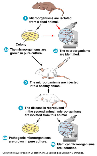
Pasteur's attempts to prove the germ theory were unsuccessful. However, the German scientist Robert Koch provided the proof by cultivating anthrax bacteria apart from any other type of organism. He then injected pure cultures of the bacilli into mice and showed that the bacilli invariably caused anthrax. The procedures used by Koch came to be known as Koch's postulates (Figure ). They provided a set of principles whereby other microorganisms could be related to other diseases.
The development of microbiology. In the late 1800s and for the first decade of the 1900s, scientists seized the opportunity to further develop the germ theory of disease as enunciated by Pasteur and proved by Koch. There emerged a Golden Age of Microbiology during which many agents of different infectious diseases were identified. Many of the etiologic agents of microbial disease were discovered during that period, leading to the ability to halt epidemics by interrupting the spread of microorganisms.
Despite the advances in microbiology, it was rarely possible to render lifesaving therapy to an infected patient. Then, after World War II, the antibiotics were introduced to medicine. The incidence of pneumonia, tuberculosis, meningitis, syphilis, and many other diseases declined with the use of antibiotics.
Work with viruses could not be effectively performed until instruments were developed to help scientists see these disease agents. In the 1940s, theelectron microscope was developed and perfected. In that decade, cultivation methods for viruses were also introduced, and the knowledge of viruses developed rapidly. With the development of vaccines in the 1950s and 1960s, such viral diseases as polio, measles, mumps, and rubella came under control.
Modern microbiology. Modern microbiology reaches into many fields of human endeavor, including the development of pharmaceutical products, the use of qualitycontrol methods in food and dairy product production, the control of diseasecausing microorganisms in consumable waters, and the industrial applications of microorganisms. Microorganisms are used to produce vitamins, amino acids, enzymes, and growth supplements. They manufacture many foods, including fermented dairy products (sour cream, yogurt, and buttermilk), as well as other fermented foods such as pickles, sauerkraut, breads, and alcoholic beverages.
One of the major areas of applied microbiology is biotechnology. In this discipline, microorganisms are used as living factories to produce pharmaceuticals that otherwise could not be manufactured. These substances include the human hormone insulin, the antiviral substance interferon, numerous bloodclotting factors and clotdissolving enzymes, and a number of vaccines. Bacteria can be reengineered to increase plant resistance to insects and frost, and biotechnology will represent a major application of microorganisms in the next century.
Image result for koch postulate

The steps of Koch's postulates used to relate a specific microorganism to a specific disease. (a) Microorganisms are observed in a sick animal and (b) cultivated in the lab. (c) The organisms are injected into a healthy animal, and (d) the animal develops the disease. (e) The organisms are observed in the sick animal and (f) reisolated in the lab.

Wednesday, April 3, 2019

The Study Of Microorganisms

The Study Of Microorganisms

As is the case in many sciences, the study of microorganisms can be divided into two generalized and sometimes overlapping categories. Whereas basic microbiology addresses questions regarding the biologyof microorganisms, applied microbiology refers to the use of microorganisms to accomplish specific objectives.

Basic microbiology

The study of the biology of microorganisms requires the use of many different procedures as well as special equipment. The biological characteristics of microorganisms can be summarized under the following categories: morphologynutritionphysiologyreproductionand growthmetabolism, pathogenesis, antigenicity, and genetic properties.


MORPHOLOGY

Morphology refers to the size, shape, and arrangement of cells. The observation of microbial cells requires not only the use of microscopesbut also the preparation of the cells in a manner appropriate for the particular kind of microscopy. During the first decades of the 20th century, the compound light microscope was the instrument commonly used in microbiology. Light microscopes have a usual magnification factor of 1000 × and a maximum useful magnification of approximately 2000 ×. Specimens can be observed either after they have been stained by one of several techniques to highlight some morphological characteristics or in living, unstained preparations as a “wet mount.”

Light microscopy

Several modifications of light microscopy are available, such as:
  • bright field
    The specimen is usually stained and observed while illuminated; useful for observation of the gross morphological features of bacteria, fungi, algae, and protozoa.
  • dark field
    The specimen is suspended in a liquid on a special slide and can be observed in a living condition; useful for determining motility of microorganisms or some special morphological characteristic such as spiral or coiled shapes.
  • fluorescence
    The specimen is stained with a fluorescent dye and then illuminated; objects that take up the fluorescent dye will “glow.”
  • phase contrast
    Special condenser lenses allow observation of living cells and differentiation of cellular structures of varying density.

Electron microscopy

The development of the electron microscope and complimentary techniques vastly increased the resolving power beyond that attainable with light microscopy. This increase is possible because the wavelengths of the electron beams are so much shorter than the wavelengths of light. Objects as small as 0.02 nm are resolvable by electron microscopy, compared with 0.25 μm—allowing, for instance, the observation of virions and viral structures. Specimens are observed by either transmission electron microscopy or scanning electron microscopy. In TEM the electron beam passes through the specimen and registers on a screen forming the image; in SEM the electron beam moves back and forth over the surface of microorganisms coated with a thin film of metal and registers a three-dimensional picture on the screen.


Advances in microscopes and microscopic techniques continue to be introduced to study cells, molecules, and even atoms. Among these are confocal microscopy, the atomic force microscope, the scanning tunneling microscope, and immunoelectron microscopy. These are particularly significant for studies of microorganisms at the molecular level.

Nutritional and physiological characteristics

Microorganisms as a group exhibit great diversity in their nutritionalrequirements and in the environmental conditions that will support their growth. No other group of living organisms comes close to matching the versatility and diversity of microbes in this respect. Some species will grow in a solution composed only of inorganic salts (one of the salts must be a compound of nitrogen) and a source of carbon dioxide (CO2); these are called autotrophs. Many, but not all, of these microbes are autotrophic via photosynthesis. Organisms requiring any other carbon source are called heterotrophs. These microbes commonly make use of carbohydrateslipids, and proteins, although many microbes can metabolize other organic compounds such as hydrocarbons. Others, particularly the fungi, are decomposers. Many species of bacteria also require specific additional nutrients such as minerals, amino acids, and vitamins. Various protozoans, fungi, and bacteria are parasites, either exclusively (obligate parasites) or with the ability to live independently (facultative parasites).
If the nutritional requirements of a microorganism are known, a chemically defined medium containing only those chemicals can be prepared. More complex media are also routinely used; these generally consist of peptone (a partially digested protein), meat extract, and sometimes yeast extract. When a solid medium is desired, agar is added to the above ingredients. Agar is a complex polysaccharide extracted from marine algae. It has several properties that make it an ideal solidifying substance for microbiological media, particularly its resistance to microbial degradation.
Microorganisms vary widely in terms of the physical conditions required for growth. For example, some are aerobes (require oxygen), some are anaerobes (grow only in the absence of oxygen), and some are facultative (they grow in either condition). Eukaryotic microbes are generally aerobic. Microorganisms that grow at temperatures below 20 °C (68 °F) are called psychrophiles; those that grow best at 20–40 °C (68–104 °F) are called mesophiles; a third group, the thermophiles, require temperatures above 40 °C. Those organisms which grow under optimally under one or more physical or chemical extremes, such as temperature, pressure, pH, or salinity, are referred to as extremophiles. Bacteria exhibit the widest range of temperature requirements. Whereas bacterial (and fungal) growth is commonly observed in food that has been refrigerated for a long period, some isolated archaea (e.g., Pyrodictium occultum and Pyrococcus woesei) grow at temperatures above 100 °C (212 °F).
Other physical conditions that affect the growth of microorganisms are acidity or basicity (pH), osmotic pressure, and hydrostatic pressure. The optimal pH for most bacteria associated with the human environment is in the neutral range near pH 7, though other species grow under extremely basic or acidic conditions. Most fungi are favoured by a slightly lower pH (5–6); protozoa require a range of pH 6.7–7.7; algae are similar to bacteria in their requirements except for the fact that they are photosynthetic.

REPRODUCATION AND GROWTH

Bacteria reproduce primarily by binary fission, an asexual process whereby a single cell divides into two. Under ideal conditions some bacterial species may divide every 10–15 minutes—a doubling of the population at these time intervals. Eukaryotic microorganisms reproduce by a variety of processes, both asexual and sexual. Some require multiple hosts or carriers (vectors) to complete their life cycles. Viruses, on the other hand, are produced by the host cell that they infect but are not capable of self-reproduction.
The study of the growth and reproduction of microorganisms requires techniques for cultivating them in pure culture in the laboratory. Data collected on the microbial population over a period of time, under controlled laboratory conditions, allow a characteristic growth curve to be constructed for a species
https://biologysciencesonline.blogspot.com/2019/04/the-study-of-microorganisms.html

TYPES OF MICROBIOLOGY,


TYPES OF MICROBIOLOGY

     
The major groups of microorganisms—namely bacteria, archaea, fungi (yeasts and molds), algae, protozoa, and viruses—are summarized below. Links to the more detailed articles on each of the major groups are provided.
Image result for type of microbiology


Bacteria (eubacteria and archaea)

Microbiology came into being largely through studies of bacteria. The experiments of Louis Pasteur in France, Robert Koch in Germany, and others in the late 1800s established the importance of microbes to humans. As stated in the Historical background section, the research of these scientists provided proof for the germ theory of disease and the germ theory of fermentation. It was in their laboratories that techniques were devised for the microscopic examination of specimens, culturing(growing) microbes in the laboratory, isolating pure cultures from mixed-culture populations, and many other laboratory manipulations. These techniques, originally used for studying bacteria, have been modified for the study of all microorganisms—hence the transition from bacteriology to microbiology.
The organisms that constitute the microbial world are characterized as either prokaryotes or eukaryotes; all bacteria are prokaryotic—that is, single-celled organisms without a membrane-bound nucleus. Their DNA(the genetic material of the cell), instead of being contained in the nucleus, exists as a long, folded thread with no specific location within the cell.
Until the late 1970s it was generally accepted that all bacteria are closely related in evolutionary development. This concept was challenged in 1977 by Carl R. Woese and coinvestigators at the University of Illinois, whose research on ribosomal RNA from a broad spectrum of living organisms established that two groups of bacteria evolved by separate pathways from a common and ancient ancestral form. This discovery resulted in the establishment of a new terminology to identify the major distinct groups of microbes—namely, the eubacteria (the traditional or “true” bacteria), the archaea (bacteria that diverged from other bacteria at an early stage of evolution and are distinct from the eubacteria), and the eukarya (the eukaryotes). Today the eubacteria are known simply as the true bacteria (or the bacteria) and form the domain Bacteria. The evolutionary relationships between various members of these three groups, however, have become uncertain, as comparisons between the DNA sequences of various microbes have revealed many puzzling similarities. As a result, the precise ancestry of today’s microbes is very difficult to resolve. Even traits thought to be characteristic of distinct taxonomic groups have unexpectedly been observed in other microbes. For example, an anaerobic ammonia-oxidizer—the “missing link” in the global nitrogen cycle—was isolated for the first time in 1999. This bacterium (an aberrant member of the order Planctomycetales) was found to have internal structures similar to eukaryotes, a cell wall with archaean traits, and a form of reproduction (budding) similar to that of yeast cells.
Bacteria have a variety of shapes, including spheres, rods, and spirals. Individual cells generally range in width from 0.5 to 5 micrometres (μm; millionths of a metre). Although unicellular, bacteria often appear in pairs, chains, tetrads (groups of four), or clusters. Some have flagella, external whiplike structures that propel the organism through liquid media; some have capsule, an external coating of the cell; some produce spores—reproductive bodies that function much as seeds do among plants. One of the major characteristics of bacteria is their reaction to the Gram stain. Depending upon the chemical and structural compositionof the cell wall, some bacteria are gram-positive, taking on the stain’s purple colour, whereas others are gram-negative.


Through a microscope the archaea look much like bacteria, but there are important differences in their chemical composition, biochemical activities, and environments. The cell walls of all true bacteria contain the chemical substance peptidoglycan, whereas the cell walls of archaeans lack this substance. Many archaeans are noted for their ability to survive unusually harsh surroundings, such as high levels of salt or acid or high temperatures. These microbes, called extremophiles, live in such places as salt flats, thermal pools, and deep-sea vents. Some are capable of a unique chemical activity—the production of methane gas from carbon dioxide and hydrogen. Methane-producing archaea live only in environments with no oxygen, such as swamp mud or the intestines of ruminants such as cattle and sheep. Collectively, this group of microorganisms exhibits tremendous diversity in the chemical changes that it brings to its environments

Algae
       The cells of eukaryotic microbes are similar to plant and animal cells in that their DNA is enclosed within a nuclear membrane, forming the nucleus. Eukaryotic microorganisms include algae, protozoa, and fungi. Collectively algae, protozoa, and some lower fungi are frequently referred to as protists (kingdom Protista, also called Protoctista); some are unicellular and others are multicellular.


Unlike bacteria, algae are eukaryotes and, like plants, contain the green pigment chlorophyll, carry out photosynthesis, and have rigid cell walls. They normally occur in moist soil and aquatic environments. These eukaryotes may be unicellular and microscopic in size or multicellular and up to 120 metres (nearly 400 feet) in length. Algae as a group also exhibit a variety of shapes. Single-celled species may be spherical, rod-shaped, club-shaped, or spindle-shaped. Some are motile. Algae that are multicellular appear in a variety of forms and degrees of complexity. Some are organized as filaments of cells attached end to end; in some species these filaments intertwine into macroscopic, plantlike bodies. Algae also occur in colonies, some of which are simple aggregations of single cells, while others contain different cell types with special functions.


Fungi

Fungi are eukaryotic organisms that, like algae, have rigid cell walls and may be either unicellular or multicellular. Some may be microscopic in size, while others form much larger structures, such as mushrooms and bracket fungi that grow in soil or on damp logs. Unlike algae, fungi do not contain chlorophyll and thus cannot carry out photosynthesis. Fungi do not ingest food but must absorb dissolved nutrients from the environment. Of the fungi classified as microorganisms, those that are multicellular and produce filamentous, microscopic structures are frequently called molds, whereas yeasts are unicellular fungi.

In molds cells are cylindrical in shape and are attached end to end to form threadlike filaments (hyphae) that may bear spores. Individually, hyphae are microscopic in size. However, when large numbers of hyphae accumulate—for example, on a slice of bread or fruit jelly—they form a fuzzy mass called a mycelium that is visible to the naked eye.
The unicellular yeasts have many forms, from spherical to egg-shaped to filamentous. Yeasts are noted for their ability to ferment carbohydrates, producing alcohol and carbon dioxide in products such as wine and bread.


Protozoa

Protozoa, or protozoans, are single-celled, eukaryotic microorganisms. Some protozoa are oval or spherical, others elongated. Still others have different shapes at different stages of the life cycle. Cells can be as small as 1 μm in diameter and as large as 2,000 μm, or 2 mm (visible without magnification). Like animal cells, protozoa lack cell walls, are able to move at some stage of their life cycle, and ingest particles of food; however, some phytoflagellate protozoa are plantlike, obtaining their energy via photosynthesis. Protozoan cells contain the typical internal structures of an animal cell. Some can swim through water by the beating action of short, hairlike appendages (cilia) or flagella. Their rapid, darting movement in a drop of pond water is evident when viewed through a microscope.


The amoebas (also amoebae) do not swim, but they can creep along surfaces by extending a portion of themselves as a pseudopod and then allowing the rest of the cell to flow into this extension. This form of locomotion is called amoeboid movement. The sporozoans (phylum Apicomplexa) are so named because they form dormant bodies called spores during one phase of their life cycle. Protozoa occur widely in nature, particularly in aquatic environments.


Viruses

Viruses, agents considered on the borderline of living organisms, are also included in the science of microbiology, come in several shapes, and are widely distributed in nature, infecting animal cells, plant cells, and microorganisms. The field of study in which they are investigated is called virology. All viruses are obligate parasites; that is, they lack metabolic machinery of their own to generate energy or to synthesize proteins, so they depend on host cells to carry out these vital functions. Once inside a cell, viruses have genes for usurping the cell’s energy-generating and protein-synthesizing systems. In addition to their intracellular form, viruses have an extracellular form that carries the viral nucleic acid from one host cell to another. In this infectious form, viruses are simply a central core of nucleic acid surrounded by a protein coat called a capsid. The capsid protects the genes outside the host cell; it also serves as a vehicle for entry into another host cell because it binds to receptors on cell surfaces. The structurally mature, infectious viral particle is called a virion.


With the electron microscope it is possible to determine the morphological characteristics of viruses. Virions generally range in size from 20 to 300 nanometres (nm; billionths of a metre). Since most viruses measure less than 150 nm, they are beyond the limit of resolution of the light microscope and are visible only by electron microscopy. By using materials of known size for comparison, microscopists can determine the size and structure of individual virions.


Prions

Even smaller than viruses, prions (pronounced “pree-ons”) are the simplest infectious agents. Like viruses they are obligate parasites, but they possess no genetic material. Although prions are merely self-perpetuating proteins, they have been implicated as the cause of various diseases, including bovine spongiform encephalopathy (mad cow disease), and are suspected of playing a role in a number of other disorders.


Lichens

Lichens represent a form of symbiosis, namely, an association of two different organisms wherein each benefits. A lichen consists of a photosynthetic microbe (an alga or a cyanobacterium) growing in an intimate association with a fungus. A simple lichen is made up of a top layer consisting of a tightly woven fungal mycelium, a middle layer where the photosynthetic microbe lives, and a bottom layer of mycelium. In this mutualistic association, the photosynthetic microbes synthesize nutrients for the fungus, and in return the fungus provides protective cover for the algae or cyanobacteria. Lichens play an important role ecologically; among other activities they are capable of transforming rock to soil.


Slime molds

The slime molds are a biological and taxonomic enigma because they are neither typical fungi nor typical protozoa. During one of their growth stages, they are protozoa-like because they lack cell walls, have amoeboid movement, and ingest particulate nutrients. During their propagative stage they form fruiting bodies and sporangia, which bear walled spores like typical fungi. Traditionally, the slime molds have been classified with the fungi. There are two groups of slime molds: the cellular slime molds and the acellular slime molds.

Mechanism of Action of Hydroxychloroquine as an Antirheumatic Drug

Mechanism of Action of Hydroxychloroquine as an Antirheumatic Drug Mechanism of Action of Hydroxychloroquine as an Antirheumat...