My Blog List

Friday, March 29, 2019

Researcher have developed a new complex hydride lithium superionic conductor that could result in all-solid-state batteries with the highest energy density

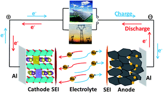
Image result for high energy sodium lithium battery
High-energy-density all-solid-state lithium metal battery employing complex hydrides.
Credit: Sangryun Kim and Shin-ichi Orimo
Scientists from Tohoku University and the High Energy Accelerator Research Organization have developed a new complex hydride lithium superionic conductor that could result in all-solid-state batteries with the highest energy density to date.
The researchers say the new material, achieved by designing structures of hydrogen clusters (complex anions), shows markedly high stability against lithium metal, which would make it the ultimate anode material for all-solid-state batteries.
All-solid-state batteries incorporating a lithium metal anode have the potential to address the energy density issues of conventional lithium-ion batteries. But until now, their use in practical cells has been limited by the high lithium ion transfer resistance, caused mainly by the instability of the solid electrolyte against lithium metal.
This new solid electrolyte that exhibit high ionic conductivity and high stability against lithium metal can therefore be a real breakthrough for all-solid-state batteries that use a lithium metal anode.
"We expect that this development will not only inspire future efforts to find lithium superionic conductors based on complex hydrides, but also open up a new trend in the field of solid electrolyte materials that may lead to the development of high-energy-density electrochemical devices," said Sangryun Kim of Shin-ichi Orimo's research group at Tohoku University.
Background
All-solid-state batteries are promising candidates for resolving the intrinsic drawbacks of current lithium-ion batteries, such as electrolyte leakage, flammability and limited energy density.
Lithium metal is widely believed to be the ultimate anode material for all-solid-state batteries because it has the highest theoretical capacity (3860 mAh g-1) and the lowest potential (-3.04 V vs. standard hydrogen electrode) among known anode materials.
Lithium-ion-conducting solid electrolytes are a key component of all-solid-state batteries because the ionic conductivity and stability of the solid electrolyte determine battery performance.
The problem is that most existing solid electrolytes have chemical/electrochemical instability and/or poor physical contact against lithium metal, inevitably causing unwanted side reactions at the interface. These side reactions result in an increase in interfacial resistance, greatly degrading battery performance during repeated cycling.
As revealed by previous studies, which proposed strategies such as alloying the lithium metal and interface modification, this degradation process is very difficult to address because its origin is the high thermodynamic reactivity of the lithium metal anode with the electrolyte.
The main challenges to using the lithium metal anode are high stability and high lithium ion conductivity of the solid electrolyte.
"Complex hydrides have received a lot of attention in addressing the problems associated with the lithium metal anode because of their outstanding chemical and electrochemical stability against the lithium metal anode," said Kim. "But because of their low ionic conductivity, using complex hydrides with the lithium metal anode have never been attempted in practical batteries. So we were very motivated to see if developing complex hydride that exhibit lithium superionic conductivity at room temperature can enable the use of lithium metal anode. And it worked."

Wednesday, March 27, 2019

Basic biology science term


acide
The acid caused corrosion. 

freeze
For water to be a solid you must freeze it. * 
oxygen
Oxygen is a gas. * 

organism
A group of living organisms consisting of similar individuals capable of exchanging genes or interbreeding. * 

alkaline
Relating to or containing an alkali; having a ph greater than 7;. * 

amphibian
A toad is an amphibian. * 

friction
The force between two moving surfaces or objects is called friction. * 

predator
Predator is a person or an animal that hunts a smaller weaker person or animal. * 
absorbThe process of something soaking, either literally or figuratively. * 
  

do violent video games promote violent behaviour in childs?

 You're under age. If you end up playing a M rate game and end up doing something stupid, it's not the responsibility of the game developer, but the responsibility of your parents who allow you to behave that way and yourself for your own actions Image result for video game images


You're 15. You're under age. You shouldn't drink, drive, watch R rated movie without adult coming with your, or play M rated video games, which is the subject of your article. 

2. You're 15!!!! You're under age. If you end up playing a M rate game and end up doing something stupid, it's not the responsibility of the game developer, but the responsibility of your parents who allow you to behave that way and yourself for your own actions. 

an image of a man in camoflauge shooting a gun in a video game environment 

3. Looks like you've been play M rate game for a while, considering some of the games you write about published a while back. If you played it when the game was first published, you're even younger. You shouldn't even be play Teen rated games, let alone M rated games. 

4. The reason there's a rating system is so that children and adolescents aren't exposed to violent material. They're brains are not ready to make decisions for themselves, or take responsibilities for their own actions. Young children can't even separate fantasy from reality. That's why we don't want kids to play Grant Theft Auto, or watch Quentin Tarantino movies. 

5. There has been studies showing a correlation between violence video games and aggression. But aggression doesn't necessarily lead to aggressive behavior. Because people are supposed to control their actions, people are supposed to restrain themselves from action upon their emotions, and when they do stupid things, they have to face the consequences. In other words, while adults might think about hurting/killing someone, most of them don't act on it. 

6. However, whether there is a correlation between violent video games and aggression doesn't matter. Even there is a correlation between violent video games and violent behavior in teenagers, it shouldn't matter because teenagers shouldn't be playing these games. It's like under age drinking, yes drinking is bad for kids, but have you heard anyone blaming vineyard? Have you heard people asking whiskey makers to only make non-alcoholic whiskeys? No. Because the responsibility doesn't lay with alcohol. It lays with the parents and liquor store who should always check ID. Young children shouldn't be playing those games, and mentally healthy adults who do playing it should have self control not to go around killing people just because they can do it in a game. 

I HATE it when people using video games as a scapegoat for bad parenting and bad behavior. For once, those parents should take the fucking responsibility for their own fucked up kids. They want video games to be their babysitter, and then blame video games for their children's behavior? Seriously? 

I once run into this nice lady in GameStop purchasing Gears of War 3 for her son, who looked no more than 10 years old. I, being a nosy person I am, asked her: you do know that it's rated M, right? She said: oh I know... but all his friends got it. All the parents of these 10 year old kids allow their children to play this M rated game in which you chainsaw enemies into bloody pieces... And these parents have the guts to blame video games to be too violent? 

new scovery Microbudibble and ultrasound radio enhancement of bladder cancer


Background

Tumour vasculature is an important component of tumour growth and survival. Recent evidence indicates tumour vasculature also has an important role in tumour radiation response. In this study, we investigated ultrasound and microbubbles to enhance the effects of radiation.

Methods:

Human bladder cancer HT-1376 xenografts in severe combined immuno-deficient mice were used. Treatments consisted of no, low and high concentrations of microbubbles and radiation doses of 0, 2 and 8 Gy in short-term and longitudinal studies. Acute response was assessed 24 h after treatment and longitudinal studies monitored tumour response weekly up to 28 days using power Doppler ultrasound imaging for a total of 9 conditions (n=90 animals).

Results:

Quantitative analysis of ultrasound data revealed reduced blood flow with ultrasound-microbubble treatments alone and further when combined with radiation. Tumours treated with microbubbles and radiation revealed enhanced cell death, vascular normalisation and areas of fibrosis. Longitudinal data demonstrated a reduced normalised vascular index and increased tumour cell death in both low and high microbubble concentrations with radiation.

Conclusion:

Our study demonstrated that ultrasound-mediated microbubble exposure can enhance radiation effects in tumours, and can lead to enhanced tumour cell death.

Main

The meshwork of blood vessels within the tumour microenvironment is constituted in part by endothelial cells that proliferate as part of the recruitment of new vessels to the tumour. These cells are critical in response to both intracellular and extracellular signals that initiate neovascularization. Anti-vascular agents that target endothelial cells are primarily focused on inhibition of angio-regulators such as vascular endothelial growth factor (VEGF), angiogenin and thrombin (Hayes et al, (1999)). Anti-vascular pharmacological agents have been successful in preclinical trials although there are several limitations to their application as a monotherapeutic approach (O’Reilly, 2006). These limitations include the requirement to administer a high dose within a prolonged treatment course in order to achieve optimal therapeutic efficacy (O’Reilly, 2006) and obstacles in targeting tumour vasculature only to spare normal vasculature in routine physiological processes (Thorpe, 2004). Phase I studies have reported toxicities associated with the administration of these pharmacological agents such as anorexia, constipation, dyspnoea, fatigue, headache, pain and hypokalemia in addition to rebound tumour growth after the cessation of therapy (Gadgeel et al, 2002Radema et al, 2002Jameson et al, 2003Rustin et al, 2003Thorpe, 2004). In the present study, we use ultrasound-mediated microbubbles as a novel treatment to perturb endothelial cells within the vascular framework in vivo.
There is a growing body of work suggesting that there can be synergistic anti-tumour effects when anti-vascular agents are combined with radiation. Recent reports propose radiation response as a function of the tumour vasculature (Raben and Ryan, 2005Nieder et al, 2006Dings et al, 2007). This is in addition to the classically accepted model that radiosensitivity and tumour control is a result of direct and indirect effects of ionising radiation on tumour stem cells and undifferentiated progenitor clonogens. Vascular damage has been long reported as an inherent effect when tumours are exposed to ionising radiation (Thomlinson, 1973). Therefore, there is an opportunity to further investigate the impact of anti-vascular agents and radiation to the tumour microenvironment.
Microbubble microspheres are coated with a lipid, protein or biopolymer, and are heavily used in medical applications of ultrasound. These microspheres encapsulate a specific gas that give them characteristic echogenicity and are currently used as contrast agents in ultrasound diagnostic imaging. Evidence that endothelial cells can be damaged during ultrasound-mediated microbubble exposure have been reported and demonstrated that endothelial cells were susceptible bystanders to injury from cavitation (Kobayashi et al, 2002). Recent investigations suggest potential in exploiting ultrasound-driven microbubbles for drug delivery in cancer therapeutics because of their affinity to permeate the endothelium of tumour microvessels (Klibanov, 2006). Advances in medical oncology are hence focusing on using microbubbles to deliver agents that would sensitise tumours to chemotherapeutic drugs by interfering with the microvasculature. Similar principles of angio-targeting are being applied to research in radiation oncology and here, we investigate microbubbles in this capacity.
A major effect caused by ultrasound interactions with microbubbles can be cavitation. Cavitation is described as rapid oscillating movements and morphological transformations that can result in outer-shell disruption of microbubbles and gas release. In cardiovascular medicine ultrasound-driven microbubbles can disrupt red blood clots, making it an effective therapy for the treatment of intravascular thrombin (Xie et al, 2005). Yu et al, 2004 reported that ultrasound-driven microbubbles could irreparably damage kidneys locally. They concluded that this modality could potentiate cell rupture from microsphere insonification in vivo (Xie et al, 2005). Previous studies have reported ultrasound-mediated microbubbles exposure to be efficacious in perturbing endothelial cells (Kobayashi et al, 2002). Endothelial cells have an important role in the vascularisation process. De novo vascularisation is reliant on the recruitment of endothelial progenitor cells that originate from the bone marrow and occur in wound healing, arthritis, inflammation and tumourigenesis (Nieder et al, 2006). Neoplastic conversion is followed by several biochemical signals that induce vascular bridging into the tumour matrix and is required for tumours to grow beyond 2 mm (Nishida et al, 2006Xuan et al, 2007). Vasculature in tumours is atypical from normal vessels resulting in haemostatic imbalance. These vessels are immature, leaky and are inefficient at supplying oxygen and nutrients (Jain, 2005Fukumura and Jain, 2007) beyond 90–140 μm. Tumour cells beyond this proximal distance adapt by modifying their activities and give rise to an acidic and hypoxic tumour environment (Cooney et al, 2006).
Power Doppler ultrasound is an important imaging tool used to investigate and map vascular structures by detecting flow of red blood cells. Power Doppler sonograms can be created by measuring echoes from moving scatterers (red blood cells that create phase shifts that are used to calculate the vascular landscape). Its advantages over other ultrasonographic imaging approaches rely on its ability to overcome common obstacles such as angle dependence, aliasing and filtering background noise (Hamper et al, 1997).
This study used high-resolution power Doppler ultrasound to monitor overall tumour response as a result of induced local damage to the tumour microvessels. We employed ultrasound-mediated microbubbles as a vascular targeting agent in combination with radiation. This study utilised a bladder cancer model as radiation can be used as a clinically definitive bladder sparing therapy and as an adjuvant modality to chemotherapy and surgery. The results of this study demonstrate that concomitant treatment with radiation and microbubbles has the ability to potentiate vascular damage by close to four-fold in certain treatment conditions in vivo. Tumours exhibited large areas of cell death caused by ultrasound-mediated microbubble exposure. The study results suggest that ultrasound-mediated microbubble treatment combined with radiation may be useful in destabilising tumour vasculature and enhancing radiation response in bladder cancer xenografts in vivo.

Materials and Methods

Cell culture

Cell lines were obtained from the American Type Culture Collection (ATCC, Manassas VA, USA). Human bladder carcinoma HT-1376 cell lines were cultured in Eagle’s minimum essential medium (ATCC) supplemented with 10% foetal bovine serum (Sigma-Aldrich, St Louis, MO, USA), 1% penicillin/streptomycin (Sigma-Aldrich) and exposed to 5% CO2 hepa-filtered air at 37 °C. Cells were cultured to 80% confluence and collected using 0.25% trypsin, 0.02% EDTA solution at room temperature. Cell pellets were isolated and re-suspended in 100 μl D-PBS (Mg−, Ca−) per 1.0 × 10⁶ cells in preparation for inducing tumours in mice.

Animals

Animal research was conducted in accordance with the guidelines by the Canadian Council on Animal Care. CB-17 white-haired severe combined immuno-deficient (SCID) male mice were obtained from Charles River Inc. (Wilmington, MA, USA). A total cell volume of 1.0 × 10⁶cells suspended in 100 μl of D-PBS (Mg−, Ca−) was injected subcutaneously to the lower right hind leg of the mouse and tumours were allowed to develop over a period of 2–3 weeks in order to reach a diameter of 5–7 mm for experiments.

In vivo image acquisition and analysis

Blood flow was detected using a VEVO770 ultrasound unit (VisualSonics, Toronto, Ontario, Canada) in power (amplitude) Doppler mode with a 25-MHz transducer (Visualsonics RMV-710B, centre frequency=20 MHz, lateral resolution=149 μm, axial resolution=54 μm, (manufacturer specifications)). The real-time microvisualization transducer was employed to acquire 3D volumetric scans (scan speed 2.0 mm s−1, wall filter 2.5 mm s−1 and step size of 0.2 mm, 90% bandwidth, 12.8 mm focal depth) where the centre of the tumour was placed at the acoustic focus. Mice were anaesthetized using a combination of 2% oxygen ventilated isoflurane for induction and then with i.p. injection of ketamine 100 mg kg−1, xylazine 5 mg kg−1 and acepromazine 1 mg kg−1 in 0.9% sodium chloride saline titrated at 0.02 ml intervals to a maximum dose of 0.1 ml. All mice were imaged before treatment administration in order to assess baseline microvessel blood flow for both acute and longitudinal studies. For ‘acute’ studies, the mice were scanned 24 h after treatment. For ‘longitudinal’ studies, the mice were scanned at 24 h and 7, 14, 21 and 28 days as established by animal health endpoints criteria. The number of images acquired per scan ranged from 60–100 frames depending on tumour size. Power Doppler images were converted into device-independent binary data for quantitative analysis using custom designed software with MATLAB (Mathworks Inc., Natick, MA, USA). Microvessel flow signals were determined by selecting a region of interest (ROI) encompassing the whole tumour in each frame. Each frame was constructed by quantised colour and non colour voxels. The vascular index (VI) was c 

Microbubbles and ultrasound activation

Definity Perflutren lipid microspheres (Lantheus Medical Imaging, North Billerica, MA, USA) were shaken using a Lantheus device for 45 s at 3000 r.p.m. Low (1% v/v) and high (3% v/v) microbubble concentrations were calculated according to total mouse blood volume estimated by animal weight. The microbubbles were diluted in sterile normal saline and injected via the tail vein. A secondary injection (0.1 cc) of normal saline was used to flush the tail vein before treatment. Mice were mounted onto a custom stage and partially immersed into a 37 °C water bath for ultrasound exposures. The ultrasound therapy system involved a micro-positioning system, waveform generator (AWG520, 

Irradiation

The tumours were X-irradiated 5 min after ultrasound treatment using an irradiation cabinet device (Faxitron, Wheeling, IL, USA). Doses of 0, 2 and 8 Gy were administered at a dose rate of 200 cGy min−1, 160 kVp energy and a source-skin distance of 30 cm as per the specifications of the device. Corporal lead sheet shielding was used with a circular aperture to expose only the tumour.

Histopathology

Tumours were fixed in 10% acetate buffered formalin (Fisher Scientific Canada, Ottawa, Ontario, Canada) following excision at 24 h (for acute studies) and 21–28 days (for long-term studies) after treatment. Tumours were fixed at room temperature for 4 h and then transferred to 4 °C for 24 h before processing using a Leica ASP300 smart tissue processor (Leica Microsystems, Richmond Hill, Ontario, Canada). Tissues were embedded in paraffin (Leica EG 1160, Leica Microsystems) and prepared as 5 μm sections onto slides with standard hematoxylin and eosin (H&E) staining techniques used for both acute and longitudinal studies. Staining using TdT-mediated dUTP-biotin nickend-labelling (TUNEL) was used in acute studies to visualise apoptotic regions. Cluster of differentiation-31 (CD31) staining was implemented to count and quantify endothelial cells within the tumour relative to the presence of intact and disrupted vessels.

Statistical analysis

A detailed analysis was conducted using GRAPHPAD INSTAT (GraphPad Software Inc., La Jolla, CA, USA) and a statistician was consulted to review the most appropriate statistical method. One-way analysis of variance statistical analysis with Dunnett’s test or Tukey’s multiple comparison’s test was performed.

Results

Microbubbles alone effectively target vasculature in HT-1376 bladder cancer xenografts in vivo

In order to quantify vasculature damage as a result of ultrasound-microbubble treatments alone, HT-1376 bladder cancer xenografts were treated with low and high concentration of microbubbles and exposed to ultrasound positioned over the tumour. The acute studies revealed that HT-1376 bladder carcinoma microvessel were responsive to treatments and demonstrated a reduction in the mean power Doppler index from the initial measured baseline. Low-concentration microbubble-ultrasound treatment after 24 h revealed a NVI was 0.91±0.01 (s.e.m.), representing a reduction in vasculature by 8.6% (Figure 1B). The higher microbubble-ultrasound treatment resulted in a NVI of 0.85±0.01 or a reduction in vasculature by 15% (Figure 1C). The long-term studies revealed similar results. Tumours that were treated with 1 and 3% microbubble concentrations when driven by ultrasound resulted in a mean power Doppler measurement of 0.71±0.01 and 0.70±0.01, respectively, after 21 days (Figures 2B and C).


Figure 1
Figure 1
Twenty four hours response monitoring of tumour vasculature using power Doppler ultrasound. (A) Tumours exposed to increasing doses of radiation demonstrated reduction in the vascularity (P<0.01, n=5). (BC) Combination therapies with low and high concentration of ultrasound-activated microbubbles and radiation. Normalised vascular index using power Doppler ultrasound was used to measure vessel abundance (P<0.01, n=5). Error bars=s.e.m


Figure 2
Figure 2
Long-term response monitoring of tumour vasculature. (A) Tumours treated with varying doses of radiation alone (P<0.001 for 0 and 8 Gy showing significant difference in vascularity; 

The combination of ultrasound-driven microbubbles and radiation synergistically decreases vasculature in HT-1376 bladder cancer tumours in vivo

Results indicated that combination treatments with microbubbles and radiation lead to synergistic anti-vascular effects as observed by power Doppler and immunohistochemistry. Short-term studies (24 h) demonstrated that at low microbubble concentration, there was a decrease in detectable flow with 2 Gy administration (NVI=0.86,±0.00) and with 8 Gy (NVI=0.73±0.00) compared with the baseline (Figure 1B). The higher microbubble concentration revealed a further decrease in detectable blood flow when combined with 2 Gy (NVI=0.79±0.01) and 8 Gy (NVI=0.66±0.00), which was greater than with microbubbles alone (Figure 1C).
Long-term studies showed similar reduction in vascularity when measured at 21 days. When 2 Gy was combined with the low microbubble concentration treatment, a NVI of 0.50±0.01 was observed and at 8 Gy with low-concentration microbubbles, a NVI of 0.43±0.00 was observed (Figure 2B). When 2 Gy radiation was combined with high-concentration microbubbles a NVI of 0.44±0.01 was observed. When 8 Gy radiation was administered with the high concentration of microbubbles in the presence of ultrasound a NVI of 0.34±0.01 was observed (Figure 2C). These changes show significant synergistic treatment effects when compared with treatment of ultrasound-driven microbubbles alone.

The combination of ultrasound-driven microbubbles and radiation significantly enhances tumour cell death in the tumour’s centre compartment caused by endothelial cell disruption

We tested to determine the efficacy of microbubbles in combination with radiation to induce vascular atrophy and to enhance tumour kill potential. We assessed tumour cell death using immunohistochemistry and measured changes in tumour size at each imaging time point. Treated tumours with combination therapy revealed tumour growth delay (Figure 3). Treated tumours demonstrated significant response to treatment within the tumour’s centre region. Circumferential tumour regions showed less significant signs of cell death according to the immunohistochemistry. Hematoxylin and eosin staining was performed on long-term studies and demonstrated an increase in cell death as higher concentrations of ultrasound-driven microbubbles and radiation were combined together (Figure 4B). Short-term treated and untreated tumours revealed insignificant changes in tumour size, however, the effects of treatment were more obvious in the long-term results. Short-term and long-term tumours showed elevated regions of cell death according to immunohistochemical (TUNEL) staining (Figure 4A).


Figure 3
Figure 3
Effects of ultrasound-activated microbubbles and radiation on the growth of HT-1376 human bladder cancer xenografts in SCID mice. Mice bearing HT-1376 human bladder xenografts were grouped for various combination treatments with ultrasound-activated microbubbles and radiation. Mice were also divided into control groups consisting of no treatment or non activated microbubbles or with ultrasound only. (A) No significant difference in tumour size for 2 Gy treatment, however, growth inhibition was observed in 8 Gy treatment with significant difference (P<0.001, n=4–5). (BC) Low and high ultrasound-activated microbubbles with varying doses of radiation showed a significant increase in tumour growth inhibition (P<0.001, n=4–5). Error bars=s.


Figure 4
Figure 4
Immunohistochemical analysis in short and long-term tumours. (A) TdT-mediated dUTP-biotin nickend-labelling staining 24 h after treatment indicates tumour necrosis resulting from local treatment. Radiation treatments alone show condensed necrotic area in multiple foci around the tumour area, which is most apparent after 8 Gy. Radiation treatments in combination with low microbubble administration exhibiting viable cells at tumour periphery. High-concentration microbubble treatments with varying doses of radiation. Severe necrotic areas observed at tumour periphery and development of cystic spaces. Scale Bar indicates 2 mm. (B) Hematoxylin and eosin staining 3–4 weeks after treatment × 20 magnification at core regions of tumours. Aberrations to the tumour microenvironment as a result of local tumour ischaemia. Top row, H&E staining through the tumour specimens demonstrated expected results with single-fraction radiation treatment of 2 Gy showed no obvious regions of cell death. Tumour cells were homogenous and evenly dispersed. With 8 Gy, cells were sparse and showed regions of decreased density, suggesting cell death. Middle row, post irradiation micropetechiae as a result of microbubble treatment response. Ultrasound-activated microbubble treatment caused a heterogeneous appearance consisting of fewer intact cells at the centre and circumscribed by healthy cells at the periphery. Bottom row, post irradiation with 3% microbubbles resulting in tumour cell necrosis at the centre. Tumours treated with combined microbubble-ultrasound and radiation demonstrated the largest areas of cell death. Bar indicates 50 μm; magnification ( × 20).


Figure 5
Long-term CD31 staining for endothelial cell markers in tumours. Cluster of differentiation-31 antibody staining was counted and quantified in tumours with varying treatments of ultrasound-activated microbubbles and radiation. (A) Cluster of differentiation-31 staining was performed for tumours 24 h after treatment. 

Discussion

In this study, we used high frequency Doppler ultrasound in conjunction with biological methods to monitor tumour response to ultrasound-driven microbubbles and radiation in a bladder cancer xenograft model. We tested the hypothesis that ultrasound-mediated microbubble treatment of tumours can be an effective vascular targeting agent that can also potentiate the effects of radiation therapy in bladder cancer xenografts in vivo. The study demonstrated that ultrasound-stimulated microbubbles in combination with radiation can induce rapid hematopoietic disruption, which presents as tumour cell death within the tumour centre in both short and long-term studies.
Microbubbles sequestered to the interior lumen of tumour vasculature can induce stress effects to the endothelium. When driven by ultrasound, microbubble disruption can result in damage to the endothelium through shear stress. This event can subsequently initiate gross vascular reorganisation within the tumour compartment. Vessel phenotypes found within the tumour compartment are typically porous, leaky, and are architecturally entangled and premature (Jain, 2005). This poses a problem in the delivery of oxygen, a major radiation enhancing molecule. In combination with cytotoxic therapies, vascular targeting agents are showing promise in tumour control by breaking the inter-reliance of cancer cells with endothelial cells in tumours  ng imaging have also utilised blood flow imaging for such studies. Goertz et al (2002) showed a reduction in doppler signals, and thus reduced blood flow perfusion within the tumour after administering an anti-vascular agent ZD126 as a monotherapy to human melanoma xenograft bearing mice. Their group demonstrated an optimal reduction in vasculature at 4 h after treatment administration and effectively presented the usefulness of Doppler sonography in monitoring vascular response. The variability in the study here has been minimised through careful handling of animals for experiments and ensuring consistent animal ages, weights and tumour sizes. Animals were treated and imaged as groups to also minimise variability. Consistency in anaesthesia and animal temperature also likely contributed to the low variability in measured Doppler signals present in this study.
Radiation treatments present several limitations in achieving optimal therapeutic outcomes because of tumour cell heterogeneity. As a result, many regimens require varying doses of radiation in order to control tumour growth and also to make a significant effect on the tumour vasculature. Our results indicated such a complication in the delivery of only 2 Gy of radiation to the tumour, demonstrating that tumour control at low doses may have weak effects on controlling the tumour, but that it can also induce pro-vascular effects through angiogenic signalling. We suspect that this induction of vascularisation through low dose radiation alone is caused by the MAP-K dependant pathway described by Park et al, 2001. Interestingly, when 2 Gy radiation was combined with low and high concentrations of ultrasound-driven microbubbles, 
Our ultrasound imaging assessment and immunohistological analysis demarcated a reduction in the vasculature and demonstrated progressive regions of apoptotic cell death within the centre of the tumour. In comparison with other anti-vascular approaches that have been shown to exhibit vascular regeneration after 7 days (Red-Horse and Ferrara, 2006), we observed no such recovery in our tumours when ultrasound-stimulated microbubbles were used in combination with radiation. Our ultrasound scans and histological analysis revealed differential vascular response around the tumour’s periphery. This circumferential vascular pattern demonstrated stable vascular abundance in contrast to the tumour’s core regions that revealed transient and sparse vasculature. We propose that this phenomenon is attributed to the vascular growth pattern before treatment. Xuan et al, 2007 used power Doppler ultrasound to detect angiogenic blood flow in prostate tumours and quantified vessel diameters of 100 μm or larger in the inner core and 12-24 μm vessels around the tumour periphery. Larger vessels feeding the centre of the tumour would result in better centre perfusion with microbubbles and may explain why vascular targeting agents tend to be well sequestered in the tumour’s core. The centre region not only showed areas of vascular atrophy but was also circumscribed by tumour necrosis. These findings may be a possible result of tumour cell starvation and a result of direct and indirect radiation targeting.

Conclusion

Vascular targeting agents can be an effective partnering modality in eradicating the tumour stroma. The current study suggests that ultrasound-mediated microbubbles can be successfully used as a vascular targeting agent and can enhance radiosensitivity in bladder cancer carcinoma. Endothelial cell disruptions and direct tumour cell kill are suspected to be implicated in both processes that render the tumour necrotic. Furthermore, this study emphasises the importance of considering multi-modality approaches in optimising the therapeutic ratio and draws attention to the importance of vascularity and endothelial cell interactions with radiosensitivity.


Mechanism of Action of Hydroxychloroquine as an Antirheumatic Drug

Mechanism of Action of Hydroxychloroquine as an Antirheumatic Drug Mechanism of Action of Hydroxychloroquine as an Antirheumat...